Cornea
Corneal Regeneration
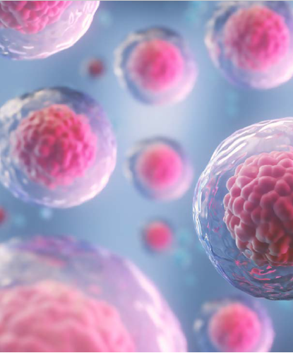
Umbilical cord stem cells a potentially limitless source of corneal endothelial cells.

Roibeard O’hEineachain
Published: Thursday, March 2, 2023
“ Improved isolation methods have resulted in reliable cultures, and we have demonstrated the potential to generate multiple corneal cell types from one piece of tissue. “
Advances in stem cell culture techniques could make human umbilical cords a valuable asset in treating corneal endothelial disease, said Jie Zhang PhD.
Dr Zhang noted in research she and her associates have carried out at the New Zealand National Eye Centre, cultured umbilical cord stem cells show promise as an alternative source for corneal endothelial cells.
“We are optimistic about the future of umbilical cord stem cells for corneal regeneration,” she said. “Improved isolation methods have resulted in reliable cultures, and we have demonstrated the potential to generate multiple corneal cell types from one piece of tissue.”
So far, their research shows umbilical cord mesenchymal stem cells and human umbilical vein endothelial stem cells can potentially differentiate into keratocytes and corneal endothelial cells. Unlike donor corneas, umbilical cords are usually discarded after birth, providing an abundant supply for use. Moreover, the cells are immunologically naïve and proliferate well.
A human option
One study led by Ms Annany Parvathi isolated umbilical cord cells obtained from Wharton’s jelly, a gelatinous substance within the umbilical cord, with all the characteristics of mesenchymal stem cells (MSCs). Flow cytometry showed the cells expressed positive markers for MSCs and no negative markers, which immunochemistry and gene expression analysis confirmed, Dr Zhang said.
When the team injected the cultured umbilical cord MSCs into the stroma of decellularised human corneal buttons, the cells migrated and repopulated the buttons. Moreover, three months after implantation, the cells expressed keratocyte genes as well as negative MSC markers, suggesting a shift away from MSC phenotype towards keratocyte phenotype.
Human umbilical vein endothelial cells (HUVECs) may provide another source for corneal endothelial cells, Dr Zhang noted. Research carried out by Ms Amnol Sandhu has developed a technique to culture a high yield of HUVECS with their characteristic cobblestone morphology and positive markers for endothelial cells.
Moreover, when cultured in conditioned media from a corneal endothelial cell line, the cells differentiated into rounder ones resembling corneal endothelial cells. They also had positive markers for zonula occludens-1 (ZO-1), a tight junction protein essential for the corneal endothelium’s barrier function.
“The results suggest HUVECs have the potential to be differentiated into corneal endothelial cells if we work on the culture a bit more,” Dr Zhang added. “Ms Sandhu is now working on identifying the factors in conditioned media that drove the differentiation and hopefully coming up with a more targeted differentiation approach.”
Five phase 2 and 3 trials using umbilical cord mesenchymal stem cells for retinitis pigmentosa and neuromyelitis optica treatment and one phase 2 trial using cord blood red blood cell concentrates for retinopathy of prematurity are underway.
“Further studies are required to determine safety and efficacy of umbilical cord stem cells for corneal disease.”
An alternative option
Yet another potential source of corneal endothelial cells are the transition zone cells from limbal rims, obtainable from corneas of donors of any age, Dr Zhang said. She noted research performed by Dr Hannah Au MBChB and Dr Jane Shi MBChB found a 62% success rate in culturing the cells. They have also found they can expand, or passage, the cultures two to six times more than endothelial cells, potentially yielding enough cells for 5,000 epithelial keratopathy patients from one donor cornea.
Transition zone cells express two essential corneal endothelial proteins, the tight junction protein ZO-1 and sodium potassium ATPase, and the neural crest protein nestin, consistent with adult stem cells for the corneal endothelium.
After three to six months in base culture, they spontaneously become endothelial-like. But to be of practical use, the cells would need to differentiate into endothelial cells at an earlier passage.
The team compared eight cell culture treatment protocols to find a speedier process for achieving the desired cell differentiation. The cell culture treatments involved combinations of Rho-kinase inhibitors and differentiation inducers.
Within seven days of treatment, the transition zone cells treated with a combination of the Rho-kinase inhibitor Y-27632 and the TGF-B inhibitor SB-431542 had rapidly become less fibroblastic in appearance compared to cells in cultures receiving other treatments or no treatment. The cells also scored highest in circularity and had reduced expression of nestin and an increased expression of corneal endothelial proteins.
“This suggests a differentiation of cells towards corneal endothelial cells, and the results suggest transition zone cells have the potential to become an alternative source of corneal endothelial cells,” Dr Zhang said.
Dr Zhang presented her paper at the 40th Congress of the ESCRS in Milan.
Jie Zhang PhD is based at the New Zealand National Eye Centre, Department of Ophthalmology, University of Auckland, New Zealand. jie.zhang@auckland.ac.nz
Latest Articles
Towards a Unified IOL Classification
The new IOL functional classification needs a strong and unified effort from surgeons, societies, and industry.
Organising for Success
Professional and personal goals drive practice ownership and operational choices.
Update on Astigmatism Analysis
Is Frugal Innovation Possible in Ophthalmology?
Improving access through financially and environmentally sustainable innovation.
iNovation Innovators Den Boosts Eye Care Pioneers
New ideas and industry, colleague, and funding contacts among the benefits.
José Güell: Trends in Cornea Treatment
Endothelial damage, cellular treatments, human tissue, and infections are key concerns on the horizon.
Making IOLs a More Personal Choice
Surgeons may prefer some IOLs for their patients, but what about for themselves?
Need to Know: Higher-Order Aberrations and Polynomials
This first instalment in a tutorial series will discuss more on the measurement and clinical implications of HOAs.
Never Go In Blind
Novel ophthalmic block simulator promises higher rates of confidence and competence in trainees.